
The Case-Coulter Translational Research Partnership, which helps to commercialize projects by clinicians and biomedical engineering faculty that improve human health and well-being, has awarded more than $1.1 million in financial backing and other support for the 2017 round of funding.
The partnership, a collaboration between Case Western Reserve University and the Wallace H. Coulter Foundation, selected seven advanced projects for full funding and four earlier-stage projects for pilot grants—research covering such patient needs as more effective brain cancer surgery, oral cancer diagnosis and devices to control pain.
Founded in 2006, the program invests more than $1.1 million annually in direct funding and support services to help research teams from Case Western Reserve advance products from the laboratory to the marketplace, where they can be available to improve patient care.
The program has contributed to 18 startup companies and several other licenses that have delivered 24 technologies to patients.
“The Case-Coulter Translational Research Partnership continues to be a cornerstone of our department, filling an essential gap to transition university biomedical technologies from research to products, where they can significantly improve the health of our society,” said Robert Kirsch, professor and chair of the Department of Biomedical Engineering.
Funding for full projects ranges from $50,000 to $200,000 per year. The money goes toward preparing projects for commercialization, such as demonstrating technical feasibility, and gauging their market feasibility and industry interest.
The program has funded more than $7.4 million in Case Western Reserve research projects since 2006, leading to more than $125 million in follow-up investment. The program directly funds about $770,000 in projects annually—a vital step in moving research from lab to real-life applications.
Projects are vetted by an external oversight committee of expert advisors from the startup community, biomedical industry and clinicians. The committee reviewed 26 technologies during the 2017 funding cycle.
“As a group, the quality of the proposals received continues to improve each year, making the selection decisions more challenging than ever,” said Stephen Fening, the program’s director. “Each year, we have more technologies worth including in the program than we can accommodate.”
Projects must have the potential to be licensed to an established or startup company within 12 to 30 months to be chosen for support.
The technologies

Novel molecular imaging agent for surgical resection of invasive brain tumors
Research leads: Susann Brady-Kalnay, professor of molecular biology and microbiology; James Basilion, professor of radiology and biomedical engineering; and Andrew Sloan, professor of neurosurgery.
Glioblastoma is a devastating brain cancer with poor survival rates because it tends to spread throughout the brain. Usually, surgery doesn’t completely remove the tumor.
The team’s novel technology is a highly selective fluorescent imaging agent, called SBK2, which could be delivered pre-surgically to cancerous tissue to make it more visible during surgery.
Being able to see the tumor in real-time is game-changing for neurosurgeons, allowing them to distinguish between normal tissue and tumor tissue to be removed. This technology could make glioblastoma surgery more effective and efficient and, eventually, surgery for other forms of cancer as well.

LunIRiS: Decision support tool for lung nodule risk prediction on screening CT
Research leads: Anant Madabhushi, the F. Alex Nason Professor II of biomedical engineering; and Robert Gilkeson, professor of radiology.
Each year, more than 20 million patients in the United States undergo a chest computer tomography (CT) exam. In nearly half of these exams, a pulmonary nodule will be identified.
While most of these nodules are benign, it is difficult to distinguish them from nodules that require treatment. As a result, many patients unnecessarily undergo more invasive diagnostic procedures, including surgical wedge resection.
The team’s novel technology, LunIRiS, is a computerized decision-support technology for use in conjunction with routine chest CT scans to reduce the high false-positive diagnostic rate associated with lung nodules. The technology could greatly reduce the number of unnecessary invasive diagnostic procedures. With advanced computational image-analytic and machine-learning tools, LunIRiS provides a risk score for improved quantitative assessment of lung nodules and has been shown to improve the diagnostic accuracy of human readers.

Point-of-care device for monitoring and diagnosis of oral cancer
Research leads: Aaron Weinberg, associate dean for research, chair of the Department of Biological Sciences and professor; Umut Gurkan, assistant professor of mechanical and aerospace engineering; and Santosh Ghosh, senior research associate.
Oral cancer kills thousands in the United States and hundreds of thousands worldwide. Early detection is key to improved survival. Oral cancer is now diagnosed by tissue biopsy, followed by pathology review.
But biopsies are expensive, painful, can cause complications and are impractical, should monitoring be required.
The team’s novel technology builds on a recent discovery that the two proteins produced in early stages of oral cancer change their ratios in cancerous cells, and that the ratio could be used as a non-invasive diagnostic tool. The researchers have developed a point-of-care microfluidic device which, when connected to a smartphone, obtains ratio results within 15 minutes.
Advantages include the ability to: non-invasively swab and diagnose a lesion for cancer while the patient waits; determine if a biopsy is necessary; permit pre-malignant lesions to be monitored; perform the test in any dental or ear, nose and throat clinic as part of oral health check-up; and obtain results at one-tenth the cost of a biopsy and pathology review.

Minimally invasive direct current nerve block
Research leads: Tina Vrabec, research assistant professor; Elias Veizi, assistant professor of anesthesiology and perioperative medicine; Niloy Bhadra, research assistant professor of biomedical engineering; Jesse Wainright, research professor of chemical engineering.
Inadequately addressed post-operative pain is a common precursor to chronic pain, which can lead to opioid addition.
The team’s novel technology uses minimally invasive direct current to block a nerve electrically. As compared to pharmaceuticals, this nerve block provides a focused block—without side effects—that can be applied and removed instantly and can be personalized for each patient.
 Polarization-sensitive OCT (PSOCT) image guidance for RFA therapy of atrial fibrillation
Research leads: Andrew Rollins, professor of biomedical engineering and medicine; and Mauricio Arruda, associate professor of medicine.
According to the American Heart Association, atrial fibrillation affects between 2.7 and 6.1 million people.
Though medical therapy is the first option, ablation therapy can be curative, although the success rate is not ideal. More than half of patients who receive ablation therapy will require additional ablation procedures, and about 35 percent will never be fully cured. Cardiac ablation requires a high level of skill and training.
Electrophysiologists don’t have real-time guidance at the catheter tip to identify critical substrates and structures to target or avoid, to assess catheter-tissue contact or to monitor the completeness of a lesion or avoid complications.
The team’s novel technology incorporates optical coherence tomography (OCT) imaging at the catheter tip, which could improve the acute success rate, reduce recurrences, procedure time and complications and improve safety.
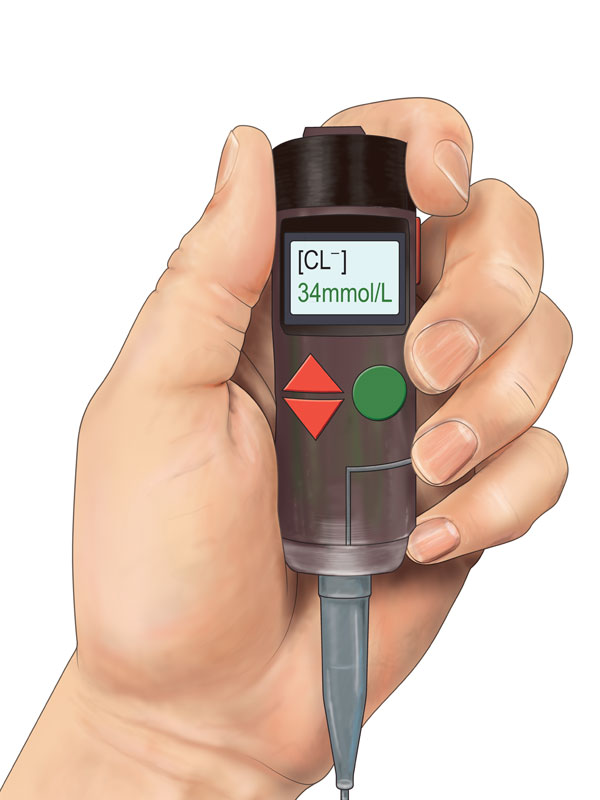
Point-of-care device for diagnosis of cystic fibrosis in newborns
Research leads: Miklos Gratzl, associate professor of biomedical engineering; and James Chmiel, associate professor of pediatrics.
Cystic fibrosis (CF) is diagnosed in one of every 2,300 live births, making it the most prevalent genetic disease in the United States, Europe and Austral-Asia.
CF is a recessive genetic disease that manifests in a defective chloride channel in epithelial cells that line internal organs. This leads to the secretion of abnormally viscous mucus with high chloride content that causes severe and life-threatening illness. The earlier CF is diagnosed, the better the patient’s quality of life, life expectancy and potential for reduced health-care costs.
The team’s novel technology promises to detect the presence of the disease in newborns at 2 weeks old, which would allow early treatment and more promising clinical outcomes.
Oropharynx appliance to maintain airway patency
Research leads: Dominique Durand, the Elmer Lincoln Lindseth Professor in Biomedical Engineering; and Kingman Strohl, professor of physiology and biophysics.
Obstructive sleep apnea (OSA) negatively impacts the health of millions of Americans, and the problem continues to grow. Sleep apnea is often cited among areas to address to reduce health-care spending and improve chronic disease management.
Non-invasive treatment options have been effective, but patients often choose to not use them because they’re uncomfortable and inconvenient.
The team’s novel technology is a device to treat OSA in a form expected to deliver much higher patient compliance. The project aims to test the new design for the treatment of OSA.
The device will be first tested in five patients for compliance in a home setting, and then undergo sleep tests at night to determine the device’s effectiveness at reducing the apnea-hypopnea index.
For more information, contact Bill Lubinger at william.lubinger@case.edu.
This article was originally published Nov. 15, 2017.