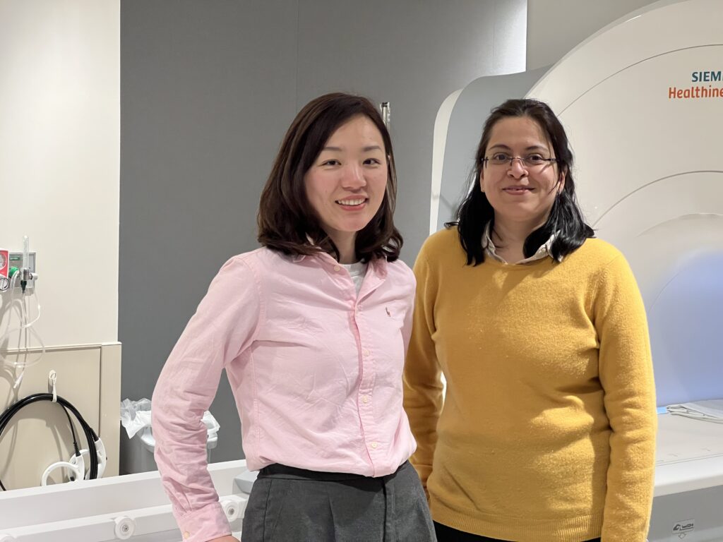
Research partnership involves academia and industry to develop and commercialize the technology
Most medical magnetic resonance imaging (MRI) scans are inherently qualitative, which can lead to subjective analysis and inconsistent conclusions.
But with a new five-year, $3.03 million grant from the National Cancer Institute—an agency of the National Institutes of Health—Case Western Reserve University researchers are leading the development and commercialization of a novel MRI and software technology that results in more accurate, consistent brain tumor diagnosis.

“Our MR scan software analyzes images quantitatively and can be reproduced,” said Dan Ma, an assistant professor in the Department of Biomedical Engineering at the Case Western Reserve School of Medicine. “To understand qualitative versus quantitative diagnosis, consider a baby with a fever. Qualitative is touching the baby’s forehead and feeling that it’s hot. Quantitative is using a thermometer for a specific temperature reading. Images from qualitative MRI scans are hard to interpret objectively, reproduce and can vary based on the type of scanners even in the same hospital.”
“This will eventually allow personalized treatment planning to improve patient outcomes,” added Chaitra Badve, a neuroradiologist at University Hospitals Cleveland Medical Center and an associate professor of radiology at the School of Medicine.
Researchers from the School of Medicine, University Hospitals Cleveland Medical Center, Siemens Healthineers and Christos Davatzikos, the Wallace T. Miller Professor of Radiology at the Perelman School of Medicine at the University of Pennsylvania, are all collaborating on this academic industrial partnership project.
The team will develop and validate a pipeline to implement the novel quantitative MR scanning technique MR Fingerprinting in the clinical workflow, and a quantitative image analysis software called MRF-QIA for MRF data analysis.
More specifically, the team is focused on demonstrating the scan and software’s effectiveness on measuring and predicting the spread of brain tumors in glioblastoma (GB) patients. GBs are highly aggressive brain tumors with a median survival of less than 15 months.
“Infiltration of cancer beyond the tumor margins causes recurrence in nearly 100% of GBs,” Badve said. “However, this cannot be measured by current MR imaging techniques, due to poor sensitivity and poor reproducibility when shared for analysis. The availability of reliable and reproducible infiltration prediction maps will lead to multi-site clinical trials in personalized radiation therapy and neurosurgery for improved patient outcomes.”
Siemens Healthineers is a market leader in medical imaging and has a nearly 40-year research collaboration with the Case Western Reserve MRI research team. The work will involve the support of a fulltime Siemens employee at CWRU and the research and development team from the company’s headquarters. Toward commercialization, Siemens will apply for U.S. Food and Drug Administration approval of the new MRF-QIA software and distribute the new analysis software for clinical implementation globally.
“We built a remarkable team of MRF developers at CWRU, image analysis and AI (artificial intelligence) experts at UPenn, clinical brain-tumor experts at UH and leading healthcare company Siemens Healthineers to ensure a successful clinical translation,” Ma said.
For more information, contact Bill Lubinger at william.lubinger@case.edu.
This article was originally published Feb. 8, 2023.

